by Stanford University Medical Center

Is SARS-CoV-2 hiding in your fat cells?
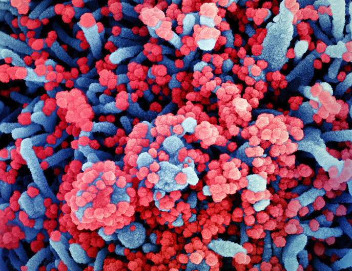
A study by Stanford Medicine investigators shows that SARS-CoV-2 can infect human fat tissue. This phenomenon was seen in laboratory experiments conducted on fat tissue excised from patients undergoing bariatric and cardiac surgeries, and later infected in a laboratory dish with SARS-CoV-2. It was further confirmed in autopsy samples from deceased COVID-19 patients.
Obesity is an established, independent risk factor for SARS-CoV-2 infection as well as for the patients’ progression, once infected, to severe disease and death. Reasons offered for this increased vulnerability range from impaired breathing resulting from the pressure of extra weight, to altered immune responsiveness in obese people.
But the new study provides a more direct reason: SARS-CoV-2, the virus that causes COVID-19, can directly infect adipose tissue (which most of us refer to as just plain “fat”). That, in turn, cooks up a cycle of viral replication within resident fat cells, or adipocytes, and causes pronounced inflammation in immune cells that hang out in fat tissue. The inflammation converts even uninfected “bystander” cells within the tissue into an inflammatory state.
“With 2 of every 3 American adults overweight and more than 4 in 10 of them obese, this is a potential cause for concern,” said Tracey McLaughlin, MD, professor of endocrinology.
The findings are described in a study published online Sept. 22 in Science Translational Medicine. McLaughlin and Catherine Blish, MD, Ph.D., professor of infectious diseases, are the study’s senior authors. Lead authorship is shared by former postdoctoral scholar Giovanny Martínez-Colón, Ph.D., and graduate student Kalani Ratnasiri.
The fat-COVID-19 connection
Obesity is defined medically as having a body mass index (weight in kilograms divided by the square of height in meters) of 30 or greater. Someone with a BMI of 25 or greater is defined as being overweight. Obese individuals are up to 10 times as likely to die from COVID-19, McLaughlin said, but increased risk for poor outcomes of SARS-CoV-2 infection begins at BMIs as low as 24.
ID-19 risk factor,” said Blish, who is the George E. and Lucy Becker Professor in Medicine. “Infected fat tissue pumps out precisely the inflammatory chemicals you see in the blood of severe COVID patients. It’s reasonable to infer that having a lot of infected fat could contribute to the overall inflammatory profile of severely ill COVID-19 patients.”
The scientists obtained samples of fat tissue from various locations in the bodies of 22 patients undergoing bariatric or cardiothoracic surgery at the Stanford Medicine Bariatric Surgery and Cardiothoracic Surgery clinic. Then, in a secure facility, the researchers infected the samples with a solution containing SARS-CoV-2 or, as a control, a SARS-CoV-2-free solution. Rigorous experiments showed that the virus could infect and replicate in fat cells, exit the cells and cause new infections in other cells.
Fat tissue contains not only fat cells but also a wide variety of immune cells, including a type called macrophages. These cells (whose name derives from two Greek words meaning “big eaters”) carry out a number of actions ranging from tissue repair and general garbage cleanup to fierce attacks on perceived pathogens—sometimes producing substantial collateral damage to normal tissue in the process.
The researchers identified a subset of macrophages in fat tissue that become infected by SARS-CoV-2, although only fleetingly. SARS-CoV-2 infection of these macrophages is abortive: It produces no viable viral progeny. But it does induce a major mood change in the macrophages.
“Once infected, these macrophages not only become inflamed themselves but also secrete substances that call in more inflammatory immune cells, in addition to inducing inflammation in uninfected neighboring ‘bystander cells,'” Blish said.
Fat tissue surrounds our hearts, guts, kidneys and pancreases, which can be adversely affected by tissue inflammation. Ominously, the scientists found infection capable of driving inflammation in virtually every SARS-CoV-2-infected fat-tissue sample they collected and analyzed.
Genetic material encoding SARS-CoV-2 was almost invariably present in fat tissue from various bodily regions of eight patients who had died of COVID-19. Examining tissue from two other deceased COVID-19 patients, the team saw an infiltration of inflammatory immune cells adjacent to infected fat cells in epicardial fat.
“This was of great concern to us, as epicardial fat lies right next to the heart muscle, with no physical barrier separating them,” McLaughlin said. “So, any inflammation there may directly affect the heart muscle or coronary arteries.”
Missing ACE2
Oddly, ACE2—the cell surface molecule that’s been implicated as the cardinal receptor for SARS-CoV-2—appeared to play little or no role in the ability of the virus to infect fat cells.
The method by which SARS-CoV-2 gains entry to fat cells and macrophages in fat tissue remains mysterious. The established primary mode of entry occurs when the virus ties up with a protein called ACE2 that sits on cell surfaces in numerous bodily tissues. Although ACE2 carries out important, legitimate functions, the virus doesn’t care what ACE2 does for a living—it considers this cell-surface protein a mere docking station.
This was the height of irony for McLaughlin and Blish, who initiated the study because they’d seen reports suggesting, although not proving, that ACE2 might be present in fat tissue. (Nobody had claimed to have sighted the protein itself, Blish added.)
But the researchers found, to their surprise, that ACE2 was virtually nonexistent on cells present in fat tissue.
“It’s highly unlikely the virus is entering through ACE2, because we couldn’t detect the functional protein in adipose tissue,” said Blish.
That means clearing SARS-CoV-2 from fat tissue could require new drugs. Monoclonal antibody therapies licensed for COVID-19, for instance, generally work by interfering with ACE2/SARS-CoV-2 interaction.
Fat tissue’s potential to serve as a reservoir where SARS-CoV-2 can hide out also raises the possibility that it could contribute to the enduring post-infection symptoms collectively called long COVID, a hypothesis that McLaughlin and Blish are beginning to explore.
Researchers from the University of Tübingen, University of Basel, Beth Israel Deaconess Medical Center in Boston and Cantonal Hospital Baselland in Liestal, Switzerland contributed to the work.
Explore further
Scientists discover a novel mechanism leading to the inflammatory cytokine storm in COVID-19
More information: Giovanny J. Martínez-Colón et al, SARS-CoV-2 infection drives an inflammatory response in human adipose tissue through infection of adipocytes and macrophages, Science Translational Medicine (2022). DOI: 10.1126/scitranslmed.abm9151 , www.science.org/doi/epdf/10.11 … scitranslmed.abm9151
Journal information: Science Translational Medicine
Provided by Stanford University Medical Center